Cavovarus Foot - High arch
- Type of Procedure: In hospital for 1-2 nights
- Length of Procedure: 3-4 hours
- Anesthesia: General anesthesia or twilight with a Nerve Block
high ARCH
What is it?
The patient with this foot deformity is characterised by having a Cavus (high arch), heel varus (heel tilted inwards) and first ray planter flexion (big toe pointing down more than normal) and forefoot pronation/adduction (front of the foot twisted inwards).
This deformity is seen in both paediatric and adult populations and in two thirds of the time is due to an underlying neurologic condition.
As a result of this not only are there bony deformities but also muscle imbalances contributing to the deformity which must be addressed.
When this is seen in both feet there is often a familial history or a congenital reason.
The patient often complains of repeated ankle sprains, pain along the outside of the foot with walking (sometimes causing stress fractures), planter callosities (thickened skin) below the first and fifth toes and heel pain as a result of planter fasciitis (inflammation of the heel).
The goal of this type of surgery is to ‘’rebalance’’ the foot and this may be achieved through osteotomies (cutting and repositioning of bones), tendon transfers and arthrodesis (fusing) of various joints if degeneration is present within the joint.
This type of surgery does not typically affect the up and down movement (dorsiflexion and plantarflexion) nor the side to side movement (inversion and eversion) of the foot and ankle.
A combination of screws and plates are inserted into the heel bone and first ray (big toe bone) to hold the bones together while the healing occurs. Once the bone is healed the hardware is not necessary but are rarely removed.
Following the reconstruction, a return to activity and exercise is important which is made easier with physical therapy and regular exercise.
In addition to this, carefully designed physical therapy program, your shoes and what you put in them are also important. An orthotic arch support which will fit in your shoe and go from shoe to shoe is important for your recovery. The orthotic support will be made by 1 of my orthotists following a computer analysis of the pressure of the foot.
General Recovery Facts
- You will not be walking on the leg without support for 8 weeks, but this depends on how quickly your bone starts to heal.
- In order to stay off your foot, you will need to use crutches, a walker, a wheelchair or a scooter type device called a roll-about.
- There will be a hard plaster cast applied to the leg for 6-8 weeks after surgery.
- Your first follow up visit will be approximately 2 weeks to inspect the wound or cast.
- If the surgery is on your left ankle, you should be able to drive an automatic vehicle at three weeks. If the surgery is on the right foot, you may drive at four weeks.
- You may begin to walk with the boot at about 6 weeks, depending upon your level of discomfort, and the instructions given to you.
- Physical therapy is helpful to regain the strength and movement of the ankle.
- You should plan to use a physical therapist for about 1-2 months.
- There will be moderate swelling of the foot, ankle and leg for about 6-9 months.
- You will continue to improve your strength and movement for about 9 months after surgery.
- Orthotic arch supports are very helpful in this recovery process.
- You may experience a variety of sensations whilst in the cast consisting of sharp shooting, dull aches, electric shocks, throbbing, sensation of itching and these are all normal and you should not panic.
- You will also be placed on DVT prophylaxis as there is a risk of blood clots shooting to the lungs with this procedure for a period of 6 weeks.
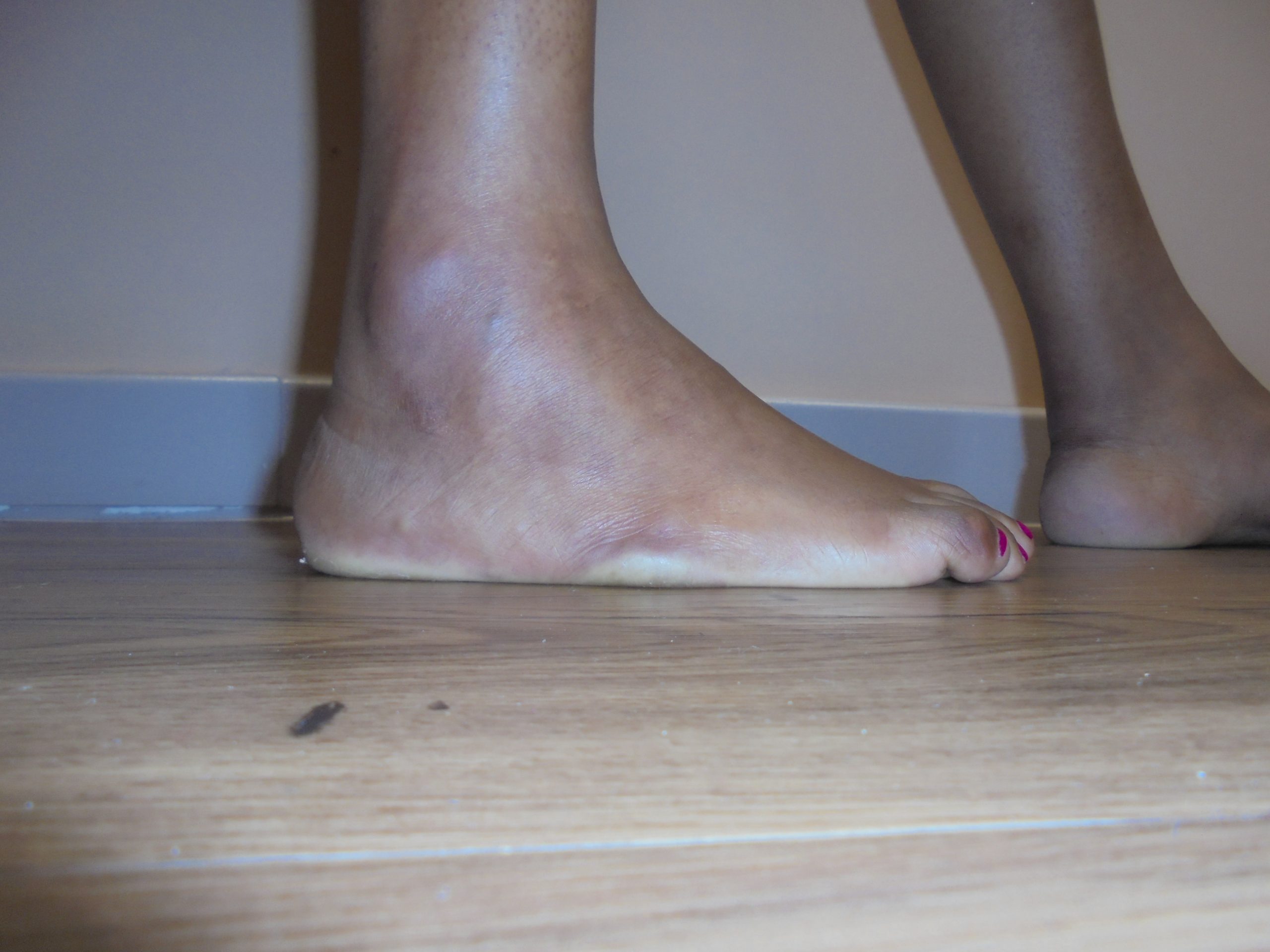
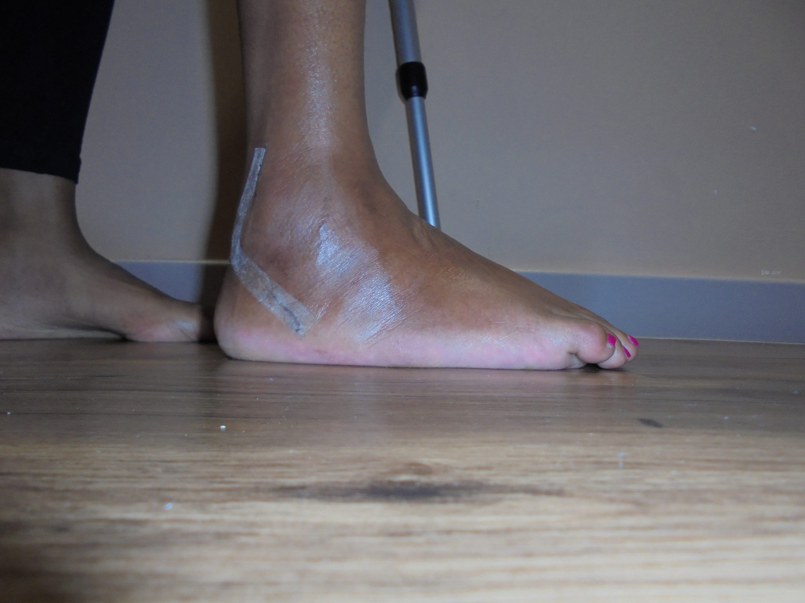
Before & After Pics
3 months post op


Specific post-operative recovery
- Foot is wrapped in a below knee splint with bandages if swelling is a concern alternatively will be placed in a below-knee cast.
- Elevate the leg on some pillows when awake however when sleeping elevate the base of the bed with 2 bricks or thick books so that the limb does not fall off the pillow at night and you wake up swollen.
- Apply ice packs.
- Take pain and DVT prophylaxis medication as prescribed.
- Expect numbness in foot 4-12 hours, followed by pain for usually one more day.
- Do not allow foot to hang down and under no circumstances bare weight.
- If you are travelling, move the toes as much as possible to stimulate the calf muscle however travel during this time is not advisable.
- First follow-up in the office.
- Wound/cast inspection for any possible complications or irritable areas.
- Application of below-knee cast for 6-8 weeks if patient previously had a splint.
- Strict non-weightbearing in cast with post-operative shoe and crutches initially to pain and swelling.
- Can allow foot to hang down at 3 weeks provided no pain and swelling occurs.
CAST REMOVED
- CT scan is taken to assess for fusion.
- Cast is removed by my dressing Sister and wound inspected.
- If incision is dry and completely healed, swimming is permitted for rehabilitation purposes taking care to not jump in the pool or apply any unnecessary force.
- Gentle exercise on bicycle or swimming in a pool with a flipper permitted preferably guided by physiotherapist.
- At this stage removable Moon boot is fitted and foot is prevented from taking excessive load and must be used for 6 weeks for any form of walking however patient does not need to sleep with this boot.
- 30lbs/13.63kgs body weight on the leg is allowed 5 minutes twice a day when washing/bathing.
- Control x-ray is taken.
- If swelling is problematic you may require the use of a compression sock.
- Start physical therapy under supervision with my protocols and this will focus on scar desensitisation and joint motion mostly.
- More vigorous exercise with Biokineticist for the next 8-12 weeks in order to achieve strength within 10% of the unaffected side hence clearing you for return to sport.
- Can discontinue boot at this stage and use a shoe with a supportive sole and a slight heel lift (heel cup) inside the sole of the shoe.
- May require gait analysis and custom orthotics to be made on an individual basis in order to assist with rehabilitation.
- Followup in my consultation rooms.
- Control x-ray taken.
- Photographs and video taken of range of motion and strength.
- Final followup in my consultation rooms.
- Control x-ray taken.
- Photographs are taken of range of motion and strength.
- Patient outcome score taken.
- Swelling and residual weakness can often be expected up until this point.
- Patient is usually discharged at this stage.